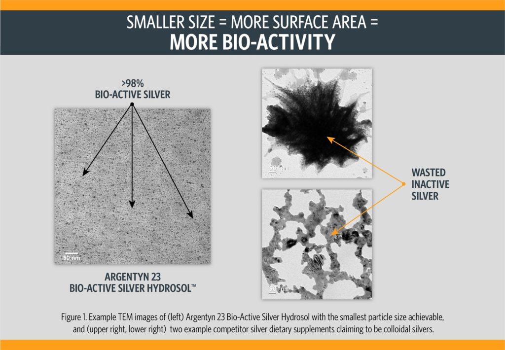
In transmission electron microscopy (TEM), silver metal appears as dark black contrast. Silver ions are too small to be imaged individually by TEM at this magnification and only contribute faint gray background patterns where they dried down. Gray contrast on TEM images can also arise from proteins, polymers, starches, and other contamination. Salt crystals appear as very uniform geometric shapes, in a range of contrast shades.
Black areas and spots in the images on the right in Figure 1 represent large silver metal particles. Compare these to the much smaller particle size and fine, uniform dispersion shown in the Argentyn 23® image on the left.
Notice that in the competitor examples shown, no individual particles are present, only large masses of silver metal. In the upper right image light gray sheets, likely from some impurities or other ingredients, are also visible in the image.
Keep in mind that bio-active silver – or useful silver – is the positively charged silver ion, and that large silver metal particles lead to wasted silver (only the outermost surface atoms of silver get converted to bio-active silver ions via surface oxidation reactions before the silver is excreted within a few hours).
Previous: Product Comparison Tool
Next: About Us


